
Soft X-Ray Spectroscopy, Microscopy, and Spectromicroscopy (WERA)
IQMT’s soft x-ray analytics facility WERA at the KIT Light Source at the Karlsruhe Research Accelerator KARA provides a coherent combination of electron spectroscopies and microscopies for studying in detail the chemical (electronic) and magnetic structure of bulk materials, thin films, and micro- and nanostructured objects.
Details (WERA)
Equipment for Electron spectroscopy and spectromicroscopy:
XAS, PES (XPS), XMCD, µ-XAS, µ-PES, µ-XMCD, topography, and NAPXAS
- Four experimental stations at WERA equipped with PEEM, electron energy analyzer, detectors, cryostats, etc.
- Sample preparation chambers, loadlocks, in-vacuo sample transfer
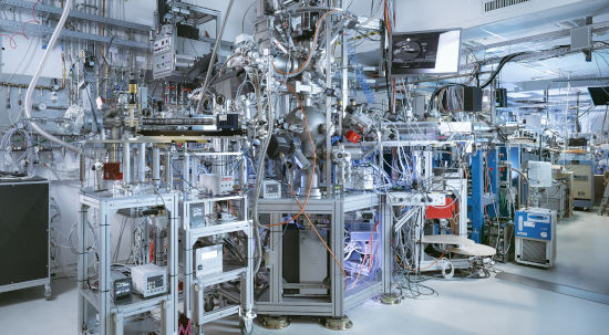
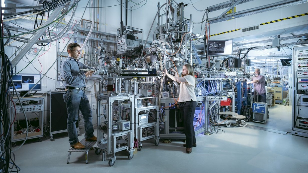
Fig. 1: Part of the cluster of experimental stations, preparation chambers, and loadlocks at WERA. (Foto: Amadeus, Bramsiepe KIT)
Features
The excitation (photon) energies are in the soft x-ray range from 100 –1500 eV, which is especially well suited for studying the light elements (like oxygen), the 3d transition metals, and the 4f rare-earth elements at their particularly informative K, L, and M edges, respectively. In the soft x-ray range, radiation-induced damage in, e.g., carbonaceous materials is orders of magnitude less than with electron bombardment (like for EELS in TEM). The photon energy resolution ΔE/E can be chosen as low as 10-4.
The main instrument for KNMFi applications is the photoemission electron microscope (PEEM). Using electron optics with great magnification, the lateral distribution of electrons emitted from the illuminated spot on the sample is imaged to an intensifier with camera readout. The lateral resolution in PEEM can be better than 30 nm (100 nm in spectromicroscopy), and this as well as the variable probing depth of <1 to about 10 nm, depending on technique, is well matched to many nano- and microstructured materials and their typical length scales. Generally, the methods are element-specific.
Two main modes are used:
Imaging of chemical (electronic), magnetic, and topographic contrast. The field of view can be chosen from 250 µm down to about 20 µm. Using the sample translation stage, a total sample area of up to about 10 mm diameter can be studied.
Spectromicroscopy: this is an especially powerful PEEM application: by taking stacks of images while tuning the photon energy or the kinetic energy of the detected electrons, laterally resolved sets of x-ray absorption (µ-XAS) and photoemission (µ-PES) spectra, resp., are efficiently obtained and can be further analyzed, see figure 2. Field of view and accessible sample area are the same as in (1). The polarization of the synchrotron radiation is a further useful variable: with circular polarization, for instance, ferromagnetic domains become visible (“magnetic dichroism”, µ-XMCD), see figure 3, and the associated spectromicroscopy gives element-specific magnetic information such as on spin and orbital magnetic moments. Linear polarization of the incident light makes the experiment sensitive to parameters like molecular and bond orientation.
In addition, WERA comprises further, complementary stations for XAS, PES (XPS), XMCD and NAPXAS. There, the signal is averaged over the illuminated spot on the sample. This allows higher energy resolution and even greater flexibility in detection methods, polarization, and sampling depth (including fluorescence detection for bulk-sensitive XAS measurements). The sample environment includes temperatures between 15 and 500 K and high magnetic field (currently 7 T).
A number of sample preparation chambers and loadlocks are part of WERA. All are interconnected with the experimental chambers by an in-vacuo sample transfer system enabling the combined investigation with methods in different chambers without the need to break the vacuum. The available preparation and characterization methods include evaporation (Knudsen cell), sputtering, annealing in vacuum or gases including oxygen, LEED. Compatibility with KARA‘s NanoLab will ensure an even wider range of possibilities for preparation and characterization.
All methods at WERA are embedded in the KNMFi user landscape. The WERA personnel has a strong commitment to user support. Please discuss your application with us.
Limitations/constraints
All experiments are performed in ultrahigh vacuum at pressures in the 10-10 mbar range. Samples should be compatible with this. (Exception: NAPXAS.)
Upgrades
WERA is constantly being upgraded and expanded. The latest upgrade includes a new experimental chamber for “near ambient pressure x-ray absorption spectroscopy” (NAPXAS) which allows investigations also on liquid samples or wet chemistry systems.
The new chamber is equipped with a fluorescence-yield (FY) detector for element-specific spectroscopy measurements and is specially designed for battery research.
The new NAPXAS chamber is currently in commissioning mode.
Typical samples

Fig. 2:
Nanolithography of phospho¬lipids by the dip-pen method (see KNMFi Laboratory for Micro- and Nanostructuring). The written triangles are 10 µm wide. The increasing admixture of nickel-chelating lipids (with a single Ni atom per lipid macro-molecule) to the carrier appears in this Ni-sensitive, spectro¬microscopic image as the increasingly red-yellow colors visible from bottom to top. Quantitative analysis reveals that for the lower four triangles, the admixture ratios observed in the written patterns directly track the original values. For the top triangle, some demixing seems to set in, leading to a smaller than-expected fraction of Ni-chelating lipids actually arriving in the written pattern. (Cf. S. Sekula et al., Small, 2008).

Fig. 3:
Magnetic domains and possibly defect-related subdomains with various orientation are visible in this magnetic dichroism image of thin film permalloy squares.
May 5, 2024
Using the new NAPXAS instrument at the Karlsruhe Research Accelerator (KARA), researchers at the Karlsruhe Institute of Technology (KIT) and the University of Münster aim to observe at the molecular level exactly how batteries charge and discharge. By making liquids accessible for synchrotron research with soft X-rays, NAPXAS provides unique insights into the processes at work in batteries.
NAPXAS (Near Ambient Pressure X-ray Absorption Spectroscopy) has been implemented at IQMT’s soft X-ray analytics facility WERA at the Karlsruhe Research Accelerator’s (KARA’s) KIT Light Source. “With NAPXAS, we can perform spectroscopic investigations of energy conversion and aging processes in batteries under more or less standard wet chemistry conditions, and we can do that live while the batteries are in use,” says Nagel, who is heading KIT’s work on the project.
After a test phase, plans call for researchers worldwide to have access to NAPXAS and WERA through the Karlsruhe Nano Micro Facility, a high-tech platform for research on functional materials in the micro- and nanometer range.
Press Release 039/2024