X-ray Absorption Spectroscopy (XAS)
XAS is an X-ray absorption spectroscopy (XAS) beamline on a dipole magnet source. Due to the high fl ux the beamline is suited for highly diluted systems in bulk samples. XAS provides the local atomic geometry (XAFS) together with electronic structure information (XANES). The information is element specific and not limited to crystalline material i.e. it is complementary to X-Ray diffraction. Distances up to 6 Å with a resolution of ±0.02 Å together with coordination number can be determined. Besides standard XAFS measured in transmission (detection limit ~5%) and fluorescence (detection limit 1 mmol/L) modes the beamline offers Q-XAFS (Quick XAFS) allowing scans as quick as 30 seconds. Grazing incidence XAFS is also possible providing surface sensitivity in the 50 nm range. A cryo cooling device for sensitive sample is available on site. At present the beamline focuses on questions in nano materials, catalytic systems and environmental science.
Contact
Dr. Stefan Mangold
Phone +49 721 608- 26073
Email stefan mangold ∂does-not-exist.kit edu
ANKA – the Synchrotron Radiation Facility at KIT
www.anka.kit.edu/english
Methods
- Extended X-ray Absorption Fine-Structure Spectroscopy (EXAFS)
- X-ray Absorption Near Edge Spectroscopy (XANES)
- Grazing Incident XAS (GI-XAFS)
Features
The XAS beamline spans the energy range from 2.4 to 27 keV. This covers the K-edges from S to Cd, and up to the L-edge of U. The double crystal monochromator design allows exchanging the two parallel mounted Si(111) and Si(311) crystal pairs within minutes. There are two principal modes of detection: transmission and fluorescence measurements. The transmission technique uses ionization chambers and the detection limit is determined by the element of interest and the other elements comprising the sample. Typical values are ~5 % by weight. The fluorescence mode is applicable for the study of elements at lower concentrations (detection limit: ~1 mmol/L) and for “thick” samples where transmission does not work. The fluorescence radiation emitted by the sample as a function of photon energy is recorded using an energy dispersive detector. Additional to standard EXAFS the XAS beamline offers grazing incident XAFS (GI-XAFS) and Q-XAFS. While the standard XAFS-mode is suited for measurement longer than 30 min, the Q-XAFS mode enables scan times of 30 sec. Even with this mode it is possible to read out the complete fluorescence spectrum for each incoming photon energy. This will enable a multi-peak fitting of the fluorescence spectra throughout the spectrum and, thus, a higher precision of the resulting data. Typical beam size is 8 mm (hor) x 1 mm (ver) (range 1 x 1 mm2 to 20 x 2 mm2).
The XAS beamline offers different types of sample stages: While the high throughput sample stage offers fast sample exchange at standard conditions, a closed cycle He-cryostat can be used for measurements down to 15 K. In future a sample stage based on cooling with liquid N2 will offer faster sample exchange with cryostat. A goniometer can be used for the integration of large set-up’s (25 kg, future 75 kg) and GI-XAS measurements.
Limitations/constraints
- Spatial resolution limited to aperture size i.e. low flux density (radiation damage is reduced - advantageous for biological specimens).
- Handling of the beamline and the data evaluation needs knowledge about XAFS.

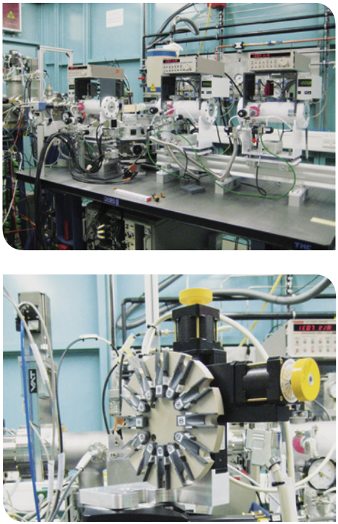
The experimental station at the XAS beamline with the goniometer as sample stage is shown in the upper picture. The picture below presents the high throughput sample stage.

